Osteopathic Institutions Reinvent Anatomy Education with XR 3D Visualization
Anatomy is a cornerstone of osteopathic education, yet today’s programs face shrinking lab time, limited cadaver access, and rising expectations for technology-enhanced learning.
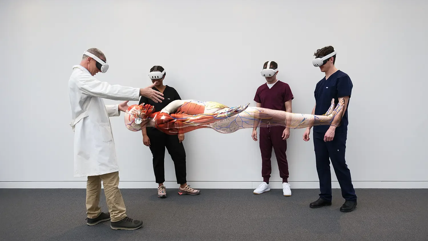
Forward-looking institutions are responding by blending tradition with immersive XR tools like HoloAnatomy® .NEXT™ to create anatomy labs that are interactive, efficient, and clinically relevant.
A.T. Still University: A Multimodal Anatomy Lab Experience

At ATSU-SOMA, anatomy was reimagined with a multimodal approach combining cadaver prosections, XR visualization, and imaging. Students rotate in small pods through each station, guided by clinical cases and quizzes on a companion app. XR reveals hidden structures with clarity through color-coding and transparency.
Faculty act as facilitators while students lead the learning, building confidence and efficiency. Exam scores improved, showing XR can strengthen student-led curricula while preserving tradition.
KansasCOM: Scaling Neuroanatomy with XR 3D Anatomy Software

At KansasCOM, XR has been central since the school’s founding. Faculty created neuroanatomy labs around a Design–Detect–Discover model, where students collaborate in pods to explore holograms of the CNS, cranial nerves, and brainstem. As enrollment grew from 90 to 200 students, XR scaled seamlessly through calibrated pods and headsets.
Learning shifted from lecture-heavy to student-led discovery guided by a companion app. Students peel back hemispheres, trace pathways, and connect anatomy directly to osteopathic manipulative medicine and clinical practice.
Insights for Osteopathic Education
Complement, don’t replace: XR and VR enhance dissection and imaging, not eliminate them.
Clinical connection: Anatomy is taught in context with imaging and clinical cases.
Faculty empowerment: Easy-to-use tools allow instructors to focus on outcomes.
Student engagement: Interactive, group-based learning sustains interest beyond the initial “wow factor.”
Why This Matters for DO Programs
For osteopathic schools, XR isn’t just an anatomy AR app, it’s a way to prepare students for practice in a healthcare system increasingly reliant on imaging, teamwork, and advanced visualization. Students enter the lab more confident, connect anatomy to clinical practice earlier, and sustain mastery in less time.
Ready to Transform Anatomy Learning at Your Institution?
Bring XR Anatomy to Your Program — Request a Guided Demo
See how institutions like A.T. Still University and KansasCOM are leading the reinvention of anatomy education. Request a guided demo of HoloAnatomy® to explore how 3D anatomy software and virtual anatomy labs can strengthen your curriculum and support your mission of preparing empathetic, practice-ready physicians.
-
HoloAnatomy is an award-winning 3D digital anatomy platform, originally developed at Case Western Reserve University’s School of Medicine. Built for mixed reality learning, it lets students and educators explore highly detailed, interactive holograms of the human body. By integrating clarity-focused 3D models with curriculum-aligned lessons, HoloAnatomy delivers a scalable, accessible alternative to cadaver dissection—designed to improve comprehension, retention, and engagement.
-
Using the Hub 3D anatomy app, instructors create customized lessons, labs, or case-based sessions that align directly with their curriculum. Learners then use XR devices (e.g., Meta Quest 3) to project life-sized anatomy into their real environment. They can walk around a holographic cadaver, zoom into organs, isolate systems, and reveal internal structures with exceptional clarity. This seamless, integrated approach functions like a virtual anatomy lab, supporting flexible teaching formats and driving better learning outcomes.
-
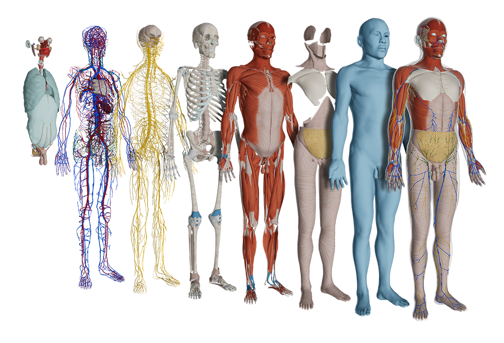
HoloAnatomy features over 7,000 meticulously crafted, color-enhanced 3D anatomy illustrations spanning 15 complete systems of the male and female human body. Developed with medical experts, the library enables exploration by system, region, organ, or custom combinations, offering comprehensive anatomical visibility for both foundational and advanced study.