Bring a New Dimension to Osteopathic Anatomy Education
Empower osteopathic medical students to build clarity, confidence, and whole-person clinical readiness through immersive 3D anatomy learning.
Partner Story:
A.T. Still University – SOMA
Founded as the world’s first osteopathic medical school, A.T. Still University’s School of Osteopathic Medicine in Arizona (SOMA) continues its tradition of innovation by integrating HoloAnatomy® into anatomy education.
Igniting student excitement: Students describe a “butterfly feeling” when first experiencing life-sized 3D anatomy, fueling collaboration and deeper engagement.
Beyond dissection: HoloAnatomy® offers interactive, reusable exploration that overcomes the cost and limitations of dissection.
Stronger understanding: Faculty and students alike highlight improved clarity of complex structures, enhancing OMM-ready learning.

Why Anatomy Matters in Osteopathic Medicine
A strong foundation in anatomy and physiology underpins every aspect of osteopathic education. Students must not only recall structures but understand how they influence function, at the heart of osteopathic philosophy. Anatomy mastery supports:
Accurate diagnosis and treatment
Confidence in performing OMM/OMT procedures
Understanding relationships between structure, function, health, and disease
Traditional methods often leave students struggling to visualize complexity. HoloAnatomy® .NEXT™ changes the game, bringing the human body to life in stunning 3D detail.
Evidence-Based Impact
Originating from Case Western Reserve University School of Medicine, HoloAnatomy® is backed by peer-reviewed research showing measurable improvements in learning:

Cut study time by up to one-third

Improve retention by up to 40%

Boost engagement & student motivation
Trusted by more than 20 leading institutions, HoloAnatomy .NEXT supports osteopathic learning environments where active, student-centered engagement drives mastery.
Tailored to your curriculum
-
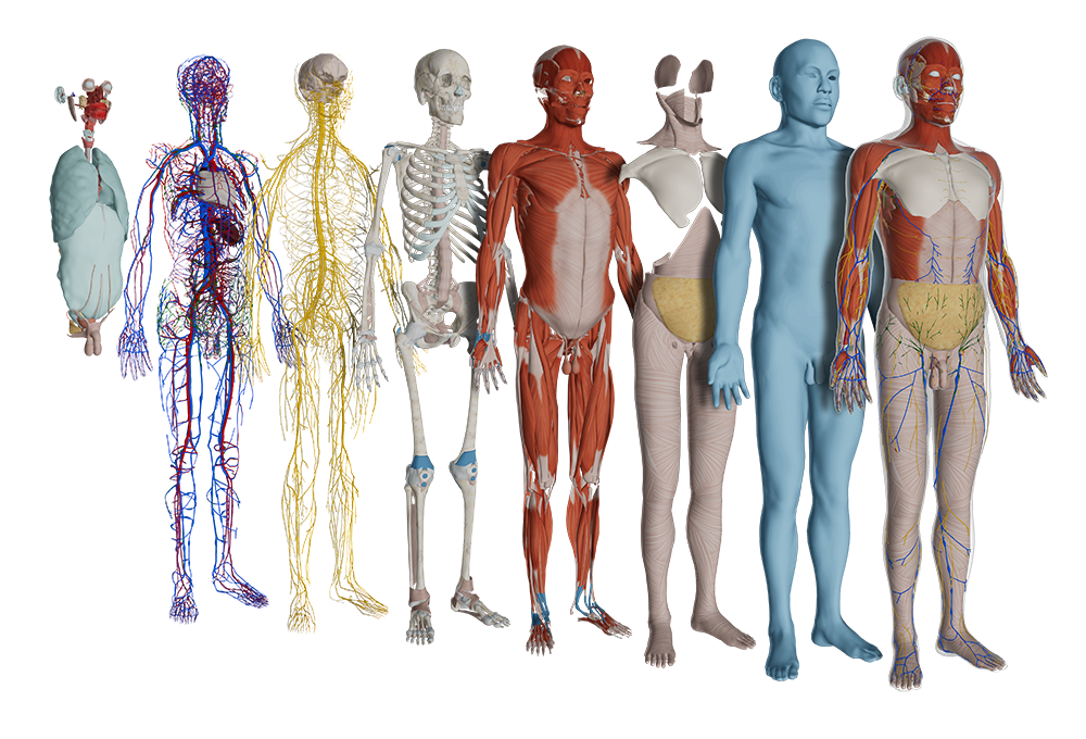
Give students clarity across every system
With detailed visibility into musculoskeletal, neurological, and vascular systems, HoloAnatomy equips students to connect anatomy to hands-on OMM practice. HoloAnatomy® Neuro further allows students gain access to the world’s most detailed 3D neuroanatomy visualizations.
-
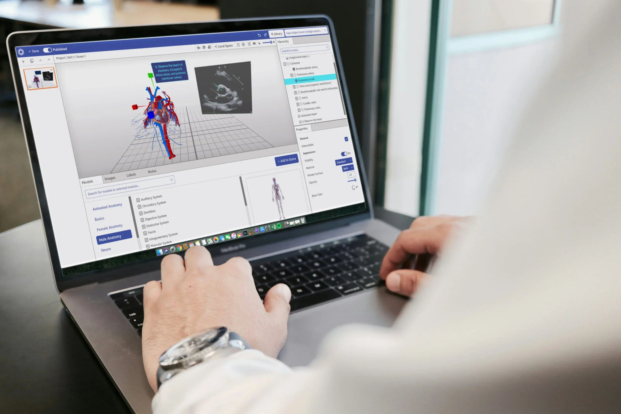
Customize lessons with Hub
From their desktop, instructors use Hub to plan sessions, integrate 3D models, images, or multimedia, and align everything seamlessly with their curriculum.
-
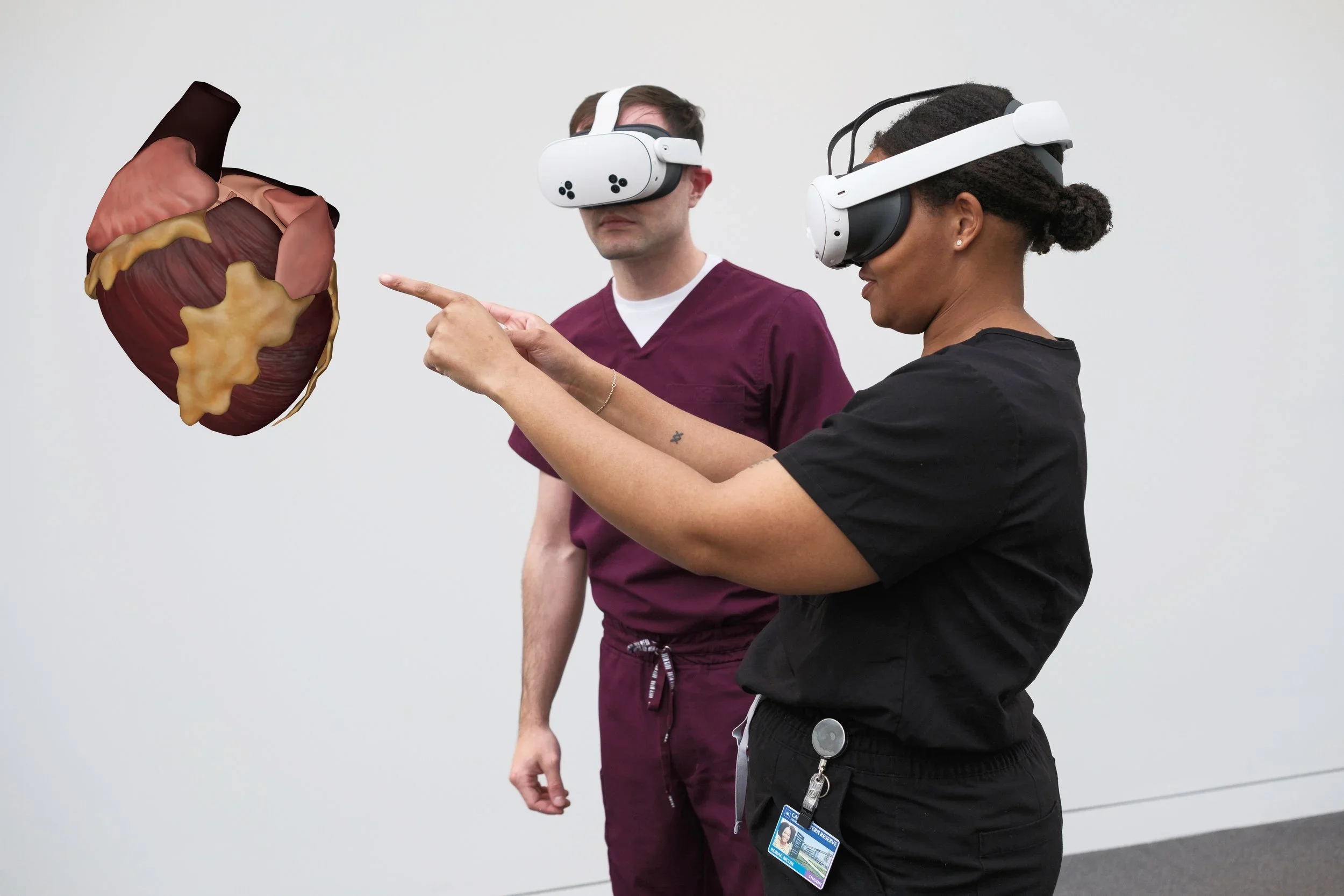
Engage students in collaborative XR learning
With Meta Quest 3 or Microsoft HoloLens, students explore anatomy at human scale, interact dynamically with structures, and collaborate with peers in real time.
Why Osteopathic Educators Choose HoloAnatomy .NEXT
Flexible integration: Tailored to medical curricula and accreditation standards
Evidence-based: Grounded in research, not just technology
Student-centered: Improves clarity, retention, and readiness for patient care
OMM-ready: Supports musculoskeletal mastery for osteopathic manipulative medicine training
Ready to reimagine anatomy for your osteopathic program?
Book a quick demonstration to explore how HoloAnatomy® .NEXT™ can strengthen your curriculum and prepare your students for whole-person patient care.
-
HoloAnatomy is an award-winning 3D digital anatomy platform, originally developed at Case Western Reserve University’s School of Medicine. Built for mixed reality learning, it lets students and educators explore highly detailed, interactive holograms of the human body. By integrating clarity-focused 3D models with curriculum-aligned lessons, HoloAnatomy delivers a scalable, accessible alternative to cadaver dissection—designed to improve comprehension, retention, and engagement.
-
Using the Hub 3D anatomy app, instructors create customized lessons, labs, or case-based sessions that align directly with their curriculum. Learners then use XR devices (e.g., Meta Quest 3) to project life-sized anatomy into their real environment. They can walk around a holographic cadaver, zoom into organs, isolate systems, and reveal internal structures with exceptional clarity. This seamless, integrated approach functions like a virtual anatomy lab, supporting flexible teaching formats and driving better learning outcomes.
-
HoloAnatomy features over 7,000 meticulously crafted, color-enhanced 3D anatomy illustrations spanning 15 complete systems of the male and female human body. Developed with medical experts, the library enables exploration by system, region, organ, or custom combinations, offering comprehensive anatomical visibility for both foundational and advanced study.