Valparaiso University Is Shaping the Future of Anatomy Learning with 3D Anatomy Software
TL;DR: Valparaiso University hosted an immersive anatomy showcase featuring HoloAnatomy® .NEXT™, showing educators how mixed reality boosts clarity, engagement, and retention, and why XR is becoming essential in health sciences education.
Valparaiso University opened its doors recently to educators and community partners for a hands-on exploration of immersive anatomy education. The event brought together faculty, students, clinicians, and regional institutions to experience how 3D anatomy software is reshaping the study of the human body, not as a distant future concept, but as a tool that students at Valparaiso are using right now.
Two regional media outlets, The Post-Tribune (Chicago Tribune) and The Times of Northwest Indiana, covered the event in depth, highlighting how Valparaiso’s Physician Assistant program is leading the way in using immersive 3D anatomy to support student understanding and confidence.
Why Anatomy Educators Came to See It Firsthand
Across disciplines, educators share a common challenge: helping students understand not just what structures exist, but how they relate, move, and function within living systems. Traditional resources including textbooks, 2D images, and dissection remain essential, but each has limitations.
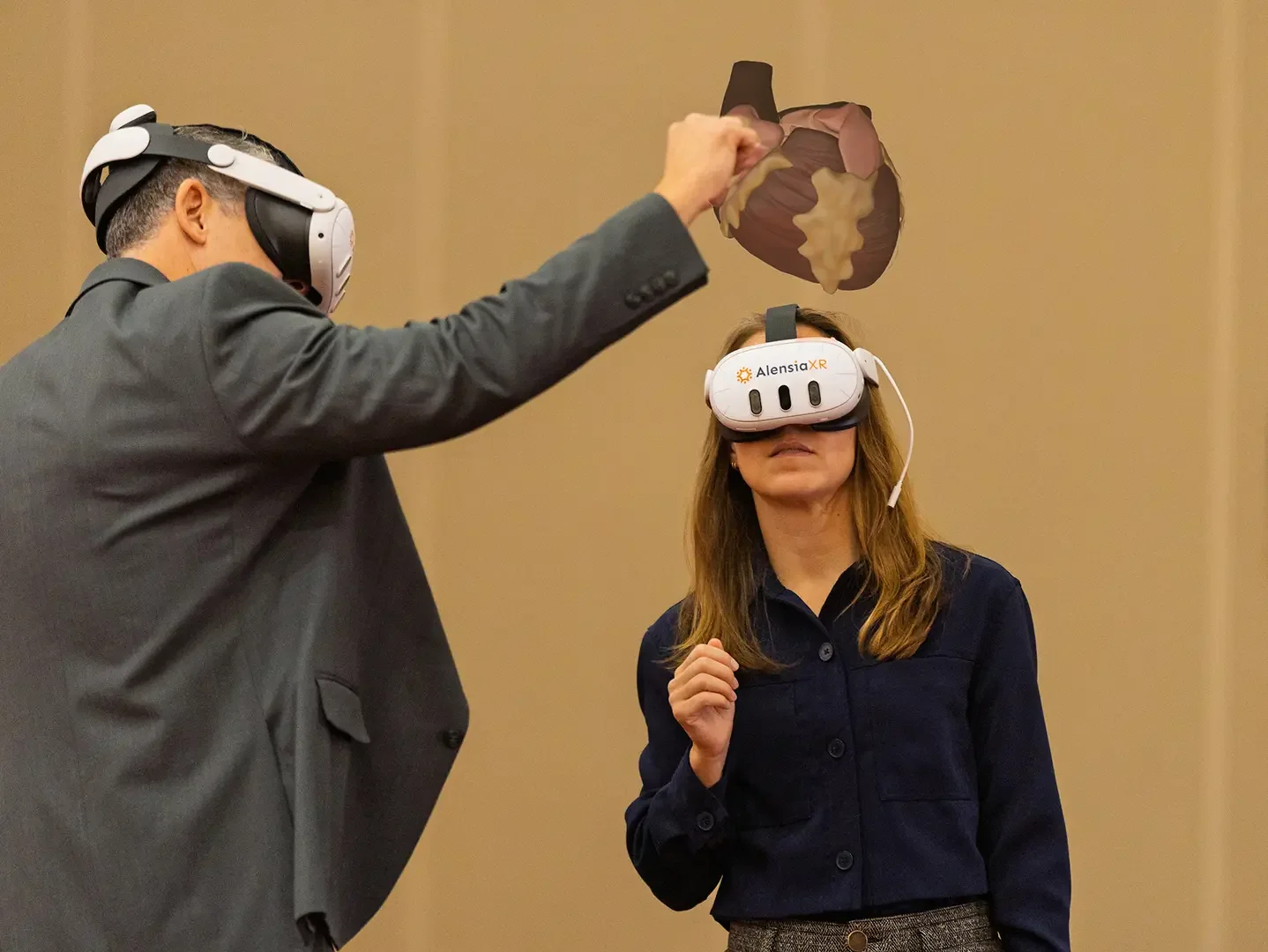
The Valparaiso Future of Anatomy Learning showcase demonstrated how extended reality (XR) adds a powerful new dimension. Participants used AlensiaXR’s HoloAnatomy® .NEXT™ platform to explore anatomical structures at true scale, interact with organ systems, and view muscle, nerve, and vessel relationships with clarity that’s difficult to achieve in any other format.
Faculty noted that this approach especially benefits visual learners and students who need to revisit content multiple times to build mastery. As one PA student shared in the Tribune article, “It’s a lot easier to actually visualize the structures.”
Key Themes and What Educators Took Away
Across dozens of conversations, three themes emerged that align strongly with the core value pillars of the HoloAnatomy .NEXT platform.
1. Flexible Curriculum Integration
Educators saw how XR can be woven directly into existing anatomy sequences, whether for introduction, reinforcement, or clinical correlation. Valparaiso’s PA program uses HoloAnatomy throughout its curriculum, enabling instructors to highlight specific structures, build scene-based lessons, and help students visualize procedures such as chest tube placement. The ability to tailor what is visible, and when, allows faculty to map immersive sessions to their exact teaching objectives.
2. Comprehensive Anatomical Visibility
The participants experienced one of the greatest strengths of the platform: clarity.
Attendees explored the shoulder, cardiac anatomy, the nervous system, and more, viewing structures at scale, examining them from multiple angles, and seeing motion in real time. The Chicago Tribune described guests “stepping inside a beating heart”, an experience that instantly communicates spatial relationships that are often abstract on paper.
This visibility helps students connect organ systems, follow pathways, and understand structure-function relationships more deeply, far beyond what traditional anatomy software or 2D images can provide.
3. Academic Origin and Evidence-Based Design
HoloAnatomy was developed in a medical education setting, and attendees saw firsthand why that matters. The content aligns with academic teaching needs, supports group interaction, and is grounded in research demonstrating improved retention, reduced study time, and stronger student engagement. Educators repeatedly mentioned how valuable it is to use a tool designed by and with medical faculty.
Looking Ahead
Valparaiso University is exploring expanded use of XR across other health sciences programs. This exploration reflects a broader shift in medical education toward virtual anatomy labs that support clearer visualization, deeper understanding, and stronger clinical connections.
For AlensiaXR, events like this affirm the growing excitement around the future of anatomy education. It’s a future where XR enhances, not replaces, the proven methods educators rely on. If you are interested in learning more about HoloAnatomy .NEXT or exploring a session for your own program, contact us and we’d be happy to help you experience it firsthand.
-
Valparaiso University’s Physician Assistant program uses HoloAnatomy® to help students visualize anatomical structures with greater clarity and understand relationships across organ systems. Faculty report that immersive 3D anatomy tools make it easier for learners to grasp complex concepts, revisit difficult regions, and build the spatial understanding needed for clinical practice.
-
Unlike static or app-based anatomy software, HoloAnatomy .NEXT provides a true virtual anatomy lab with real-time 3D visualization, motion-enhanced models, and collaborative exploration. Educators can tailor lessons, highlight structures progressively, and integrate clinical correlations, supporting more flexible curriculum design and more effective learning outcomes.
-
Mixed Reality creates a virtual anatomy lab where students can collaborate while exploring structures at scale, see motion in real time, and revisit difficult regions for better mastery. When XR anatomy education complements traditional methods, institutions commonly report clearer understanding, higher retention, and more engaged learners.