Reimagining Neuroanatomy: How KansasCOM is Leading the Way with Immersive Technology

Kansas College of Osteopathic Medicine
Kansas College of Osteopathic Medicine is pioneering a new era in medical education by reimagining neuroanatomy instruction without relying on cadavers. Their forward-thinking approach, combined with the rich history of progressiveness in Kansas, led them to explore immersive technology as a way to enhance student learning and engagement. AlensiaXR sat down with Dr. Cameron Jeter, Professor and Chair, Department of Biomedical Sciences, to learn about their journey with HoloAnatomy® Neuro.

AlensiaXR: How was neuroanatomy taught before considering immersive technology?
Dr. Jeter: The school decided some time ago to teach anatomy without relying on donors and dissection. We explored various methods to supplement traditional textbook learning, like using arts and crafts to mimic neuroanatomy structures. While engaging, methods like Styrofoam cups and colored yarn weren’t scalable. We needed technology that worked for us. As someone born and raised in Kansas, it was crucial for me to uphold our tradition of leadership and progress while pioneering neuroanatomy education.
AlensiaXR: What led you to consider HoloAnatomy® Neuro?
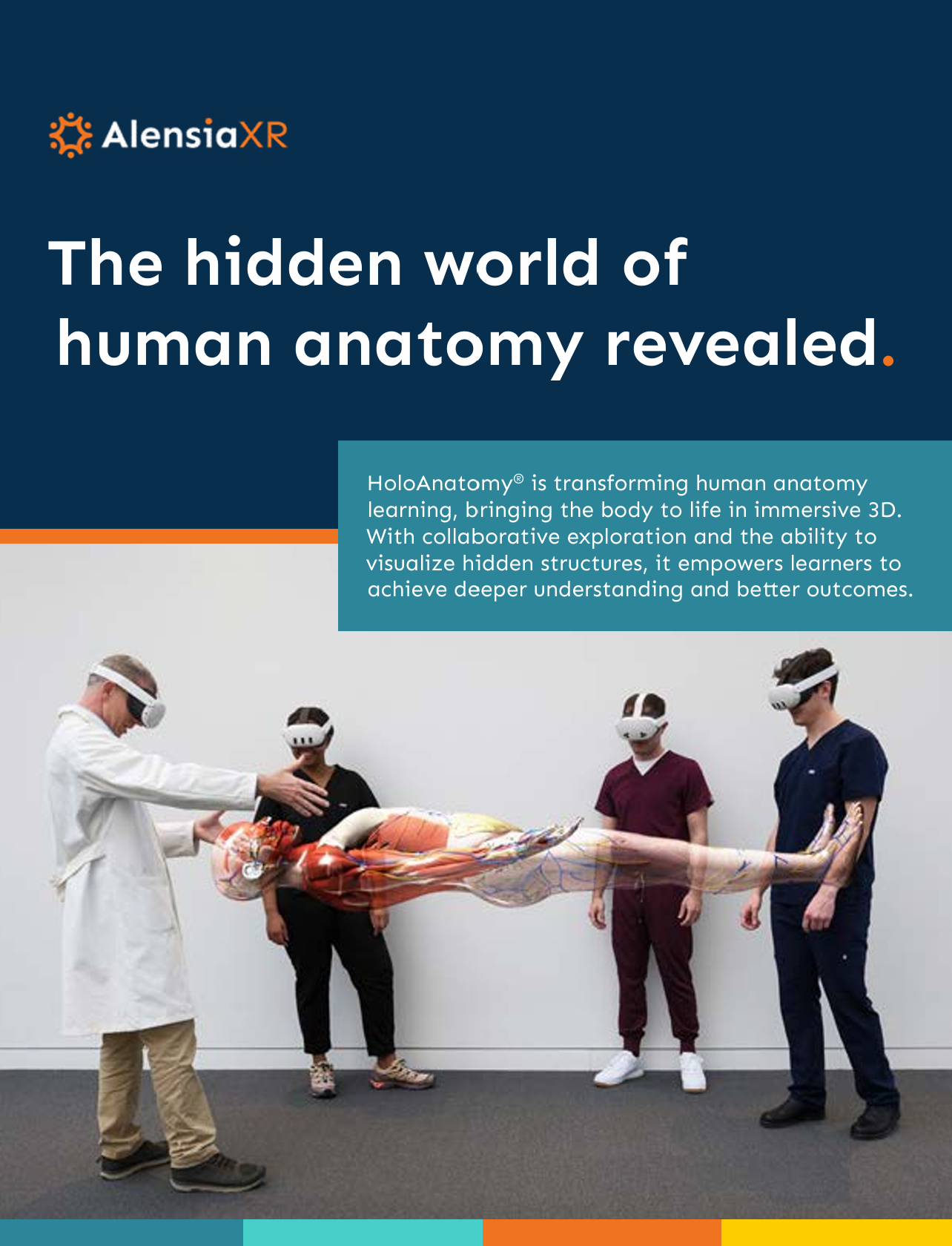
Dr. Jeter: We explored the pros and cons of multiple technologies. Initially there was hesitance, but we quickly realized that we needed to revisit the top options. We wanted more than viewing 3D images on 2D displays merely mimicking the immersive experience. We wanted to be able to explore cross-sections, spinal cords, and brain stems. We wanted to be selective of which structures to show. It was important, for example, to help students visualize how a neuron travels through the spinal cord to the muscle.
AlensiaXR: What has been your experience deploying & using the solution?
Dr. Jeter: One thing is clear – we could not have done it without a team. AlensiaXR provided the training to faculty, and our AV team was key to helping us deploy. A key contributor to our success was a Medical Education Specialist who was both tech savvy and knowledgeable about neuroanatomy. Faculty visualized the flow of instruction with storyboards, articulating to the specialist what they wanted to see on the holographic slides. For example, they might want to show the cortical spinal tract for a lesson. Our specialist made the selections to assemble the slides, shared a draft for feedback, then we went to the lab to experience the results.
AlensiaXR: How did faculty and students react to being immersed in neuroanatomy?
Dr. Jeter: Everyone experienced a “Wow” moment when using Neuro for the first time. Being able to view microscopic tracts as a rainbow of structures was exciting. Even student’s body language changed. Some of them would actually step back in surprise and awe. There would be silence as they took it in, a lot of talking. Once they had the overcome the “wow moment”, the curiosity came in. If there was any initial hesitation, it melted away as they interacted with the holograms and each other.

AlensiaXR: Have you observed any benefits from using HoloAnatomy® Neuro?
Dr. Jeter: In addition to students being more engaged, we found students actually taking ownership of the learning process. Students can work in small groups or pods, with each member having an equal role. We provide worksheets to focus learning, and the students seamlessly dive right in. Compared to cadaver study, the prep time and the clean-up time is nearly zero while avoiding the costs of cadavers.
Even with traditional dissection, organs are not always in their proper anatomical space because they have dropped or moved or shifted, whereas with Neuro they are in their true anatomical position, occupying the space where they should.
Download the HoloAnatomy eBook
Learn about its origin, transformative growth, and invaluable benefits experienced by a growing list of institutions and organizations.
